FIGURE 1:
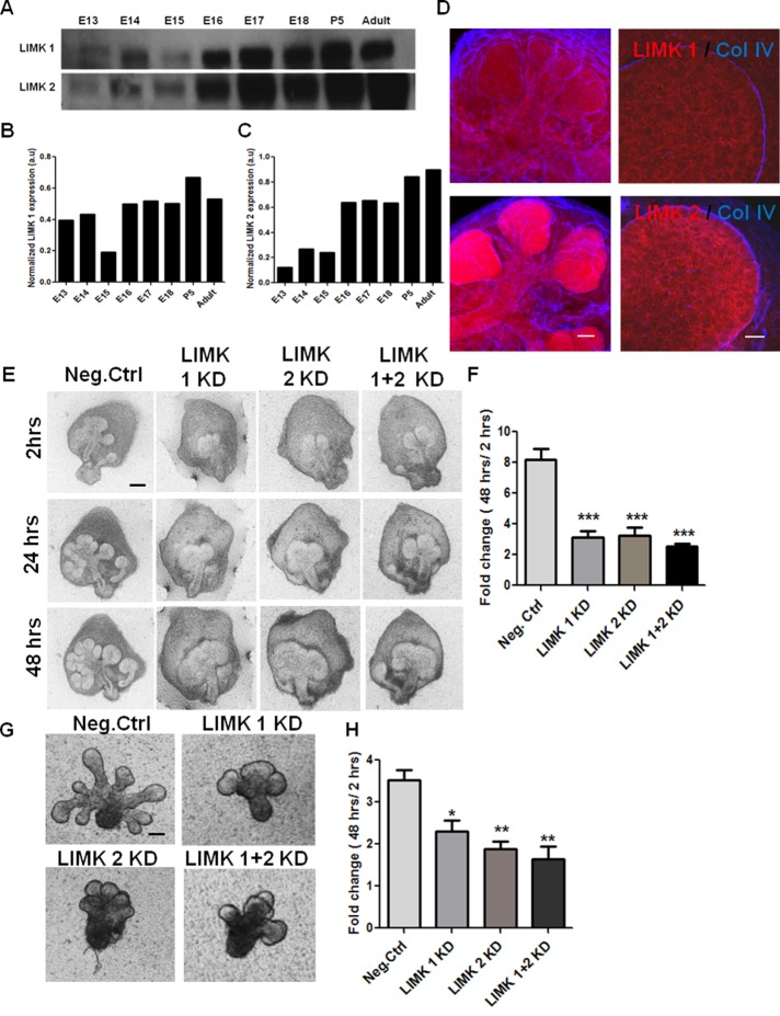
LIMK 1 and 2 are required during early development of salivary glands. (A) Western blot analysis and densitometric quantification of total SMG protein extracts shows LIMK 1 and 2 isoforms to be present in the embryonic stages E13–E18, postnatal day 5 (P5), and adult. (B, C) LIMK 1 and 2 protein expression levels quantified relative to GAPDH levels, respectively. (D) E13 SMG organ explants were grown for 24 h and subjected to ICC/confocal imaging. LIMK 1 (red, top) is in both epithelium and mesenchyme, outside the basement membrane, as indicated by collagen IV (blue). LIMK 2 (red, bottom) is found primarily in the epithelium. Scale, 50 μm (left, 20×), 20 μm (right, 63×). (E) E13 SMGs were treated with LIMK1, 2, or 1 + 2 siRNA and subjected to bright-field imaging. Scale, 200 μm. (F) Morphometric analysis shows a significant reduction in bud number (fold change 48 h/2 h) with LIMK 1, 2, or 1 + 2 siRNA relative to negative control (n = 15). (G) Mesenchyme-free E13 epithelial rudiments treated with LIMK 1, 2, or 1 + 2 siRNA also show a decrease in branching after 48 h. Scale, 200 μm. (H) Morphometric analysis of fold changes in bud number reveals a reduction in completed buds in LIMK 1, 2, or 1 + 2 siRNA–treated glands at 48 h (n = 10). *p < 0.05, **p < 0.01, ***p < 0.001, ANOVA.

