FIGURE 10:
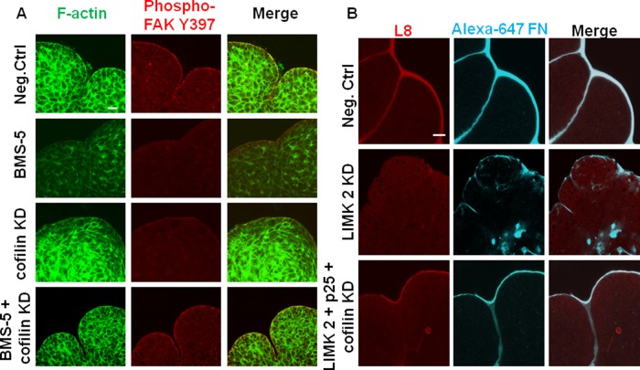
Rescue of LIMK inhibition restores F-actin dynamics along with focal adhesion kinase activity and FN deposition and assembly in the cleft regions of embryonic glands. (A) Mesenchyme-free E13 epithelial rudiments treated with BMS-5, cofilin siRNA, or BMS-5 plus cofilin siRNA for 24 h were subjected to ICC/confocal imaging to detect pFAK (Y397, red) vs. F-actin (green). The decreased F-actin with LIMK inhibition by BMS-5 and increased F-actin with cofilin siRNA both disrupted FAK activation in the cleft region. Cofilin KD in the presence of BMS-5 stabilized actin dynamics and restored downstream FAK activation. Scale, 20 μm. (B) Mesenchyme-free E13 epithelial rudiments treated with control siRNA or LIMK2 ± cofilin and p25 siRNAs for 24 h were grown in the presence of Alexa 647–labeled human plasma FN (cyan) and subjected to ICC/confocal imaging to detect assembled fibrillar FN (L8 antibody, red). The decrease in assembled FN (L8- red) around the epithelial periphery relative to negative control–treated SMGs and aberrant accumulation of FN (cyan) observed in the interior of the epithelium in LIMK 2 siRNA–treated glands is rescued with simultaneous addition of cofilin and p25 siRNA. Scale, 20 μm.

