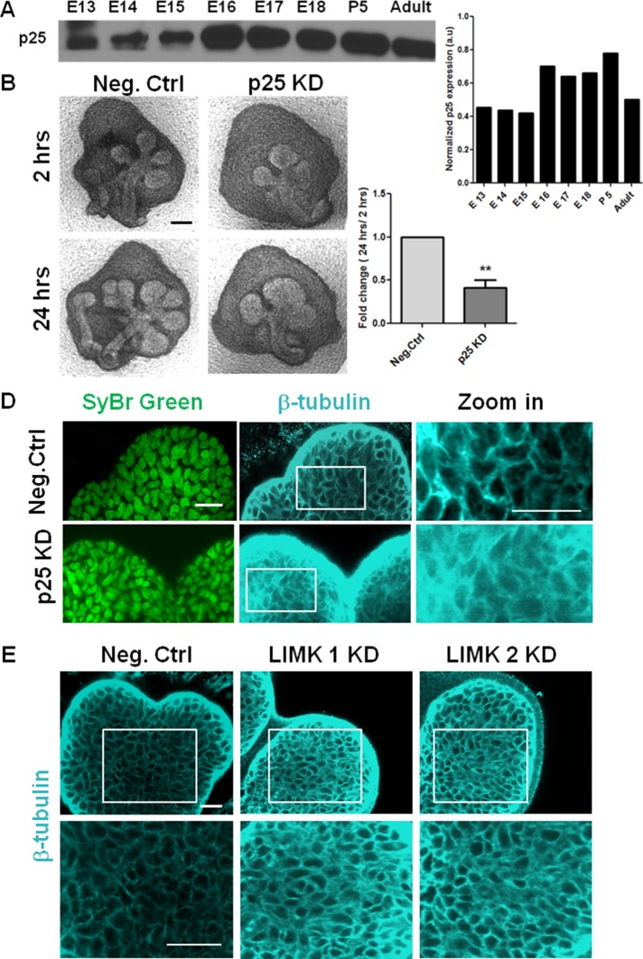
FIGURE 3:
Regulation of microtubule dynamics by TPPP/p25 and LIMK is required during branching morphogenesis. (A) Western blot analysis and densitometric quantification show developmental expression of p25 protein from E13 to E18, P5, and adult. (B) E13 SMG organ explants were grown for 24 h ± p25 siRNA and imaged with bright-field microscopy. Scale, 200 μm. (C) Morphometric analysis shows a significant reduction in buds with p25 KD; n = 10, paired t test, **p < 0.01. (D) Mesenchyme-free E13 SMG rudiments were grown for 24 h ± p25 siRNA, stained for SyBr green (green), and subjected to ICC for β-tubulin (cyan) and imaged with confocal microscopy. In p25 KD, β-tubulin exhibits a diffuse distribution in the epithelium relative to negative control siRNA–treated glands (as seen in zoomed-in image), indicative of reduced microtubule stability. Scale, 20 μm. (E) Mesenchyme-free E13 SMG rudiments were grown for 48 h ± LIMK 1 or 2 siRNA vs. negative control siRNA. LIMK 1 and 2 KD demonstrated increased β-tubulin staining at the cell borders (bottom, zoomed-in area) in the epithelium relative to negative control siRNA–treated glands, indicative of overstabilized microtubules. Scale, 20 μm.

