FIGURE 1:
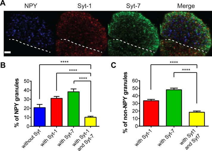
Confocal imaging of Syt isoform expression on NPY-containing granules. (A) Chromaffin cells cultured on glass coverslips were transfected with a plasmid encoding NPY-Cer. At 3–4 d after transfection, cells were fixed, permeabilized, and exposed to Syt-1 and Syt-7 antibodies. Confocal sections of 0.5 μm were taken through the cell. The region closest to the coverglass was imaged. The dotted line indicates the boundary of the transfected NPY-Cer cell from a nontransfected cell below. Scale bar, 3 μm. (B) The percentage colocalization of NPY granules with any synaptotagmin or with Syt-1, Syt-7, and Syt-1 plus Syt-7. Differences between groups were assessed with the Student's t test (n = 17 cells, ****p < 0.0001). (C) Synaptotagmin isoforms were sometimes detected in regions without obvious NPY-Cer fluorescence. Colocalization of isoforms in non-NPY granules was determined. Differences between groups were assessed with the Student's t test (n = 17 cells, ****p < 0.0001).

