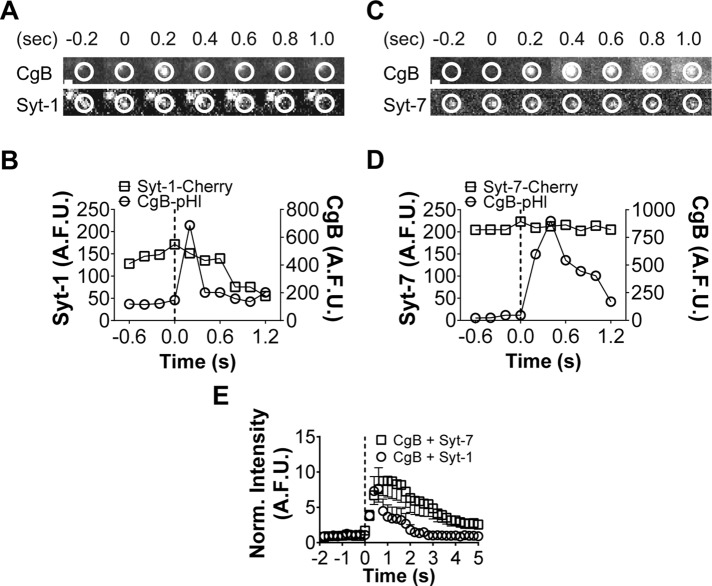
FIGURE 7:
Syt-1 granules release lumenal contents more quickly than Syt-7 granules. (A) Fusion of a Syt-1-Cherry granule. CgB-pHluorin intensity increases (indicating fusion) at time 0.2 s (within circled region). Syt-1 and CgB disperse quickly after fusion (dotted line). Note loss of fluorescence within circled region (A, B). (C) Syt-7 remains punctate after fusion (bottom). CgB is released slowly. (D) Graphs for C. (E) On average, CgB is more rapidly released from fusing Syt-1 rather than Syt-7 granules. n = 19 for Syt-1 plus CgB; n = 21 for Syt-7 plus CgB. After 2 s, averages ± SEM are significantly (*p < 0.05) different by Student's t test. Scale bar, 960 nm.

