FIGURE 6:
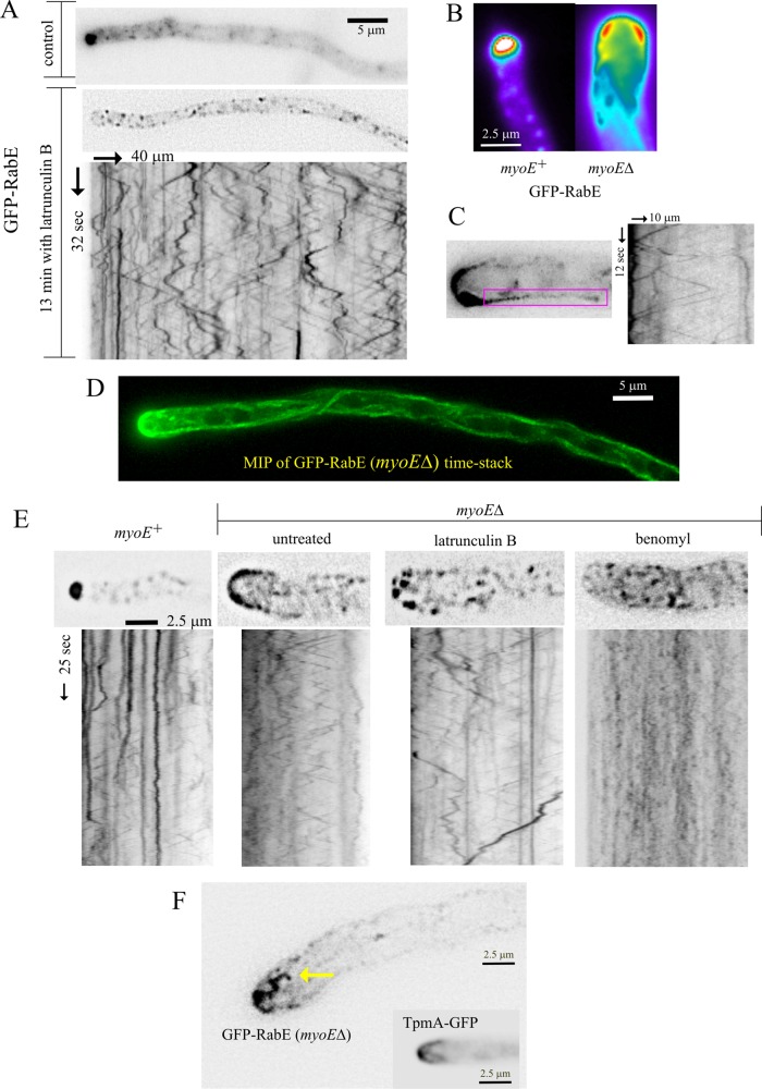
MyoE-dependent and -independent functions of F-actin. (A) Latrunculin B treatment delocalizes GFP-RabE–containing membranes at the SPK to the MT conveyor belt. Derived from Supplemental Movie S8. (B) An example of a myoE∆ tip in which the GFP-RabE crescent is thicker in a slightly subapical position. (C) Kymograph covering the position of a cortical MT, showing that it serves as track for RabERAB11 carriers moving in both directions and the uniform speeds characteristic of MT-dependent movements. (D) Maximal-intensity projection of a middle-plane time stack showing how moving GFP-RabE carriers delineate trajectories of MTs. (E) Micrographs from time stacks of images and the corresponding kymographs, showing effects of F-actin and MT depolymerization on the tip-and-apical crescent-associated GFP-RabE carriers. Both drugs delocalized these carriers from the tips of myoE∆ cells, but these carriers moved only when MTs were present. (F) Isolated frame from a time stack (Supplemental Movie S11) showing microfilament-like material decorated with GFP-RabE carriers. A hyphal tip in which actin cables are decorated with GFP-tropomyosin is shown for comparison. Experiments were carried out with strain MAD4120 and its myoE∆ derivative, MAD4409. The TpmA-GFP inset corresponds to MAD1750.

