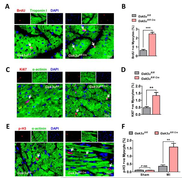
Figure 5. Deletion of Gsk3α promotes post-MI cardiomyocyte proliferation.
(A) Representative images show BrdU-positive cardiomyocytes from the WT and Gsk3α KO hearts. (B) Quantification shows significantly increased numbers of BrdU-positive myocytes in 3 wk post-MI KO hearts, n=6 for each group. (C) Images show Ki67 positive cardiomyocytes. (D) Quantification shows an increased number of Ki67-positive cardiomyocytes in Gsk3α KO hearts, n=4-5. (E) Representative images show p-H3 (Ser10)-positive myocytes in 3 wk post-MI hearts. (F) Quantification shows an increased number of p-H3 positive myocytes in the KO hearts, n=4 for sham and n=6 for MI group. Results are expressed as mean±SEM. ** P<0.01, *** P<0.0001.

