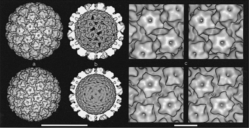
Figure 3.
Surface-shaded representations of CRPV (top row) and HPV-1 (bottom row) reconstructions viewed along a 2-fold symmetry axis. The CRPV capsid has an “open” form and the HPV-1 has a “closed” form. a, Outside view of virions. b, Inside view of capsids (back half). Chromatin cores were computationally removed from the virion density map (radii < 46 nm for CRPV and < 42 nm for HPV-1). c, Closeup stereo views. In CRPV (top), elongated holes lie on opposite sides of the 2-fold axis. Triangular holes on the 3-fold axes can be seen at the top and bottom center, just behind the protruding point of the capsomere. In HPV-1 (bottom), there is a single, small hole on the 2-fold axis. Bars represent: a,b, 50 nm; c, 10 nm.

