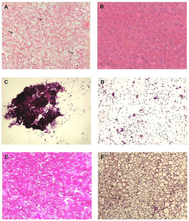
Fig. 2.
Histological analysis of livers from LS-G6pc−/− and control mice. Paraffin-embedded H&E-stained liver sections from a 2-month-old LS-G6pc−/− mouse (A) and control mouse (B). Arrows show macro- and micro-lipid vesicles in the enlarged hepatocytes. Liver fine-needle aspiration cytology sample stained with PAS from a 2-day-old LS-G6pc−/− mouse (C) and a 2-day-old control mouse (D). Paraffin-embedded liver sections from 3-month-old LS-G6pc−/− mice were stained with PAS (E) or PAS plus diastase (F). (A,B,E,F) 10× magnification; (C,D) 20× magnification.

