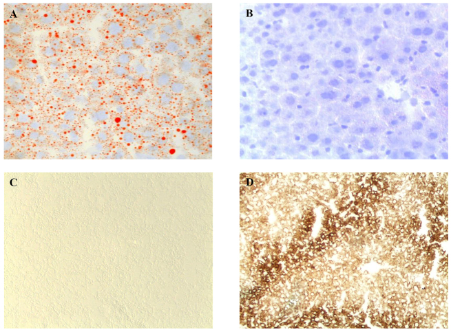
Fig. 3.
Histochemical analyses of liver from LS-G6pc−/− mice. Oil red O staining of LS-G6pc−/− mouse (A) or control mouse (B) liver cryostat sections at 2 months of age shows lipid accumulation in the hepatocytes of the LS-G6pc−/− mouse. To evaluate G6Pase activity, liver cryostat sections were treated as described in the Materials and Methods. Colored lead sulfide was developed with ammonium sulfide. Representative results of liver sections from 3-month-old LS-G6pc−/− (C) and control (D) mice are shown. (A,B) 20× magnification; (C,D) 10× magnification.

