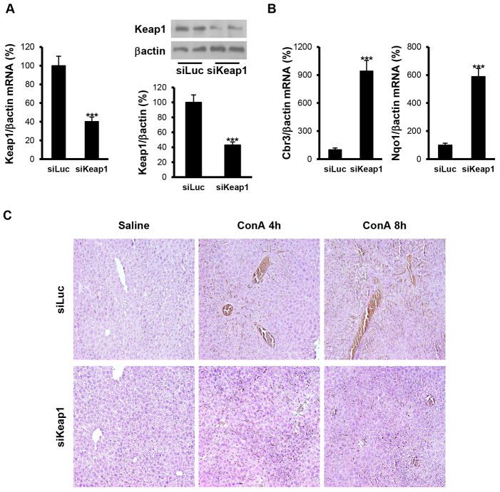
Fig. 1.
Reduction of Keap1 in the liver by siRNA administration in vivo attenuates ConA-induced liver damage. Analysis of Keap1 expression and histopathological features in liver samples from luciferase siRNA (siLuc) or Keap1 siRNA (siKeap1) mice after 4 and 8 hours of ConA treatment (n=4–6 animals per condition). (A) (left panel) Keap1 mRNA levels determined by real-time PCR. (Right panel) Representative blots with the indicated antibodies and quantification of the densitometric analysis from all blots. Data are presented as mean±s.e.m. relative to siLuc mice. (B) Cbr3 and Nqo1 mRNA levels determined by real-time PCR. Data are presented as mean±s.e.m. relative to siLuc mice. (C) Representative images from hematoxylin and eosin staining. ***P<0.005, siKeap1 versus siLuc.

