Fig. 6.
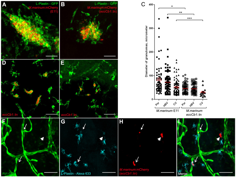
Role of the ESX-1 locus in the formation of early brain granulomas. (A) Example of a compact cluster with phagocytic cells and bacteria after infection into the parenchyma with M. marinum E11 (red), stained with anti-L-plastin (green). (B) Example of cluster after infection into the parenchyma with M. marinum eccCb1::tn (red), stained with anti-L-plastin (green). (C) Overview of diameter of individual granulomas counted in embryos infected via parenchyma (Par), hindbrain ventricle (HBV) or caudal vein (CV), with either M. marinum E11 or the isogenic eccCb1::tn mutant. The number of granulomas is described in Table 1. Par E11 versus Par F3.1: *P<0.0001, HBV E11 versus HBV F3.1: **P<0.0001, CV E11 versus CV F3.1: ***P=0.0135. (D) Example of single phagocytic cells filled with mycobacteria in an embryo infected into the parenchyma with eccCb1::tn mutant (red), stained with anti-L-plastin (green). (E) Example of single phagocytic cells filled with mycobacteria in an embryo infected via the caudal vein with eccCb1::tn mutant (red), stained with anti-L-plastin (green). Scale bars: 35 μm (A,B,D,E). (F–I) Z-stack of the relationship between vasculature, phagocytes and bacteria. Caudal vein infection with M. marinum eccCb1::tn mutant, with (F) Tg(Fli1:GFP)y1 casper embryo showing blood vessels, (G) L-plastin Alexa-Fluor-633 showing phagocytic cells and (H) eccCb1::mcherry showing bacteria. (I) Merge of panels F–H. Closed arrow indicates a phagocyte containing mycobacteria inside a blood vessel; open arrow indicates a phagocyte containing mycobacteria outside a blood vessel; arrowhead shows a bacterial cluster probably inside a dying phagocyte. Scale bars: 50 μm (F–I).

