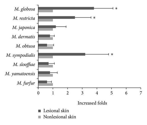
Figure 3.

The relative quantity of each Malassezia species in the lesions compared with the nonlesional skin of the 24 patients. The y-axis displays the increased prevalence of the species in the lesions compared with the nonlesional skin. ∗P < 0.05, nonlesional skin versus lesional skin.
