
Figure 1.
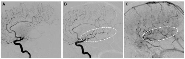
Example of Modified Thrombolysis in Cerebral Infarction 2a (mTICI 2a). Lateral projection images of M1 occlusion at baseline (A), post-treatment early arterial (B), and late parenchymal-venous (C) phases demonstrate restoration of antegrade flow into the inferior division branches, which produces a capillary blush in <50% of the middle cerebral artery territory (dotted ovals).