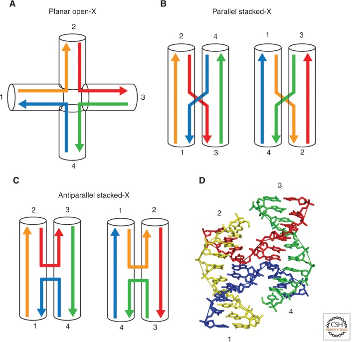
Figure 2.
Global conformations of DNA Holliday junctions. The component helices are drawn in different colors and labeled 1 to 4 in a clockwise manner. (A) In the absence of divalent cations, four-way junctions adopt a fully extended conformation with no coaxial stacking between the component helices (planar open-X form). Electrostatic repulsion between phosphates keeps the junction in an unstacked conformation with the four arms directed toward the corners of a square. (B,C) In the presence of divalent cations, the helices undergo coaxial stacking such that the symmetry is lowered from fourfold in the fully extended form to twofold in the stacked forms. Pairwise coaxial stacking can occur in two different ways, characterized by the stacking of helices 2 on 1 and 3 on 4 (left) or 1 on 4 and 3 on 2 (right). The stacked X junction can adopt either parallel (B), or antiparallel (C) conformations. (D) Crystal structure of a Holliday junction (Eichman et al. 2000) in the right-handed antiparallel stacked-X conformation (Protein Data Bank, PDB, ID 1dcw), showing the continuous strands in yellow and green and exchanging strands in red and blue.

