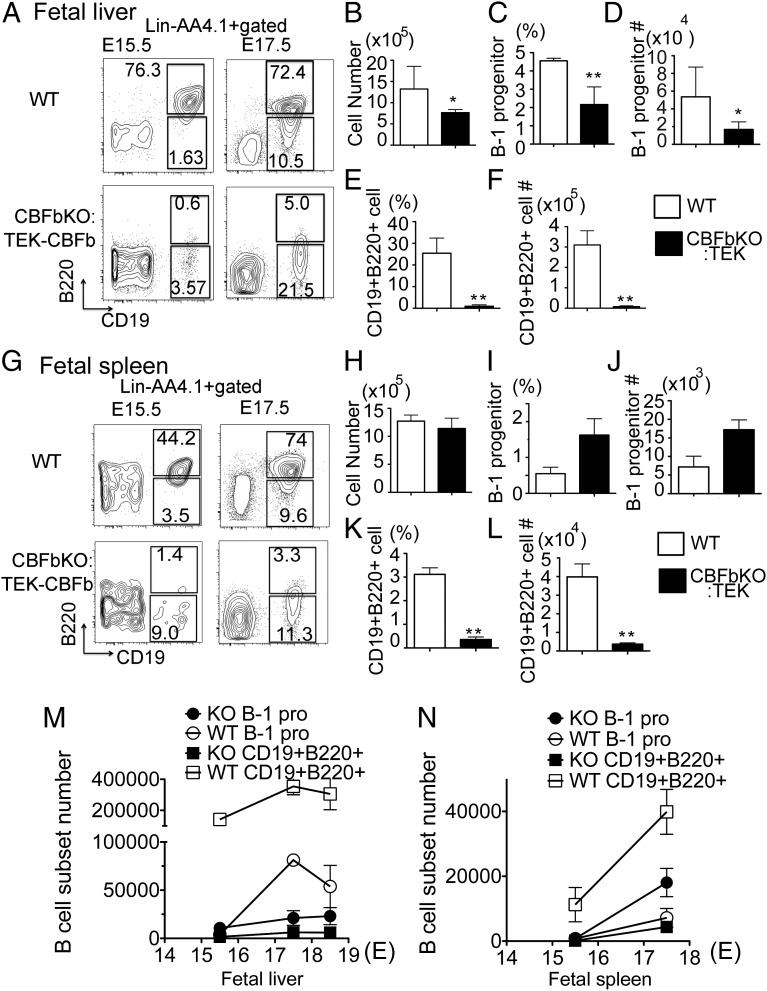
Fig. 1.
B-1 progenitor cells exist in the Cbfβ−/−:Tek-GFP/Cbfβ fetal liver (FL) and spleen. AA4.1+CD19+B220lo-neg B-1 progenitor phenotype was examined in the WT and Cbfβ−/−:Tek-GFP/Cbfβ fetal-liver and spleen cells at each day from E15.5 until birth (E18.5). The representative data for E15.5 and E17.5 are depicted. Lin−AA4.1+ gated FACS dot plots at E15.5 and E17.5 fetal liver (A) and fetal spleen (G) are depicted. Total MNCs of E17.5 FL (B) and spleen (H), percentage of B-1 progenitor cells in FL (C) and spleen (I), total cell number of B-1 progenitor cells in FL (D) and spleen (J), percentage of AA4.1+CD19+B220+ cells in FL (E) and spleen (K), and total cell number of AA4.1+CD19+B220+ cells in FL (F) and spleen (L) are shown. (B–F and H–L) Open bar, WT; filled bar, Cbfβ−/−:Tek-GFP/Cbfβ. The cell numbers of B-1 progenitors and CD19+B220+ cells from WT and Cbfβ−/−:Tek-GFP/Cbfβ FL (M) and spleen (N) at each embryonic day are shown (n = 4 for each group; *<0.05, **<0.01).

