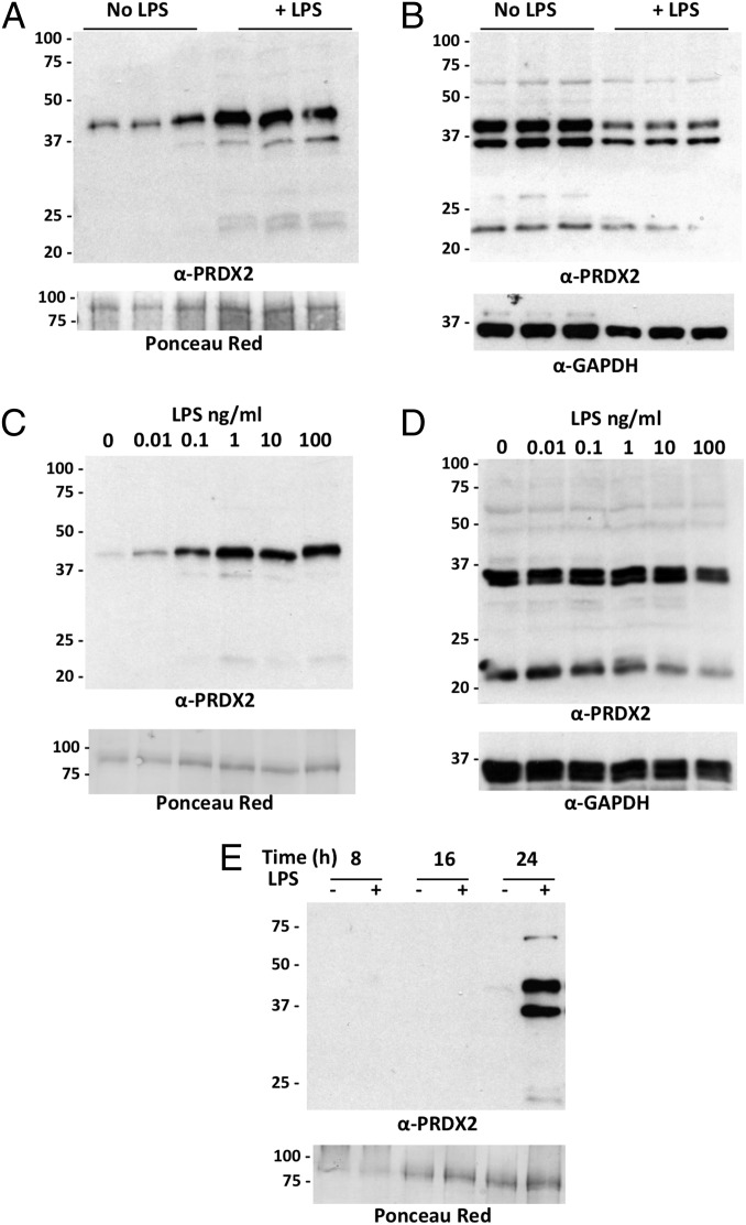
Fig. 2.
LPS induces PRDX2 release. (A) Western blot of supernatants from RAW264.7 cells treated for 24 h with and without 100 ng/mL LPS. PRDX2 was measured in triplicate samples each derived from an independent well. (Upper) Western blot with anti-PRDX2. (Lower) Ponceau red staining. (B) Intracellular PRDX2 in the cell lysates of the experiment in A. (Upper) Western blot with anti-PRDX2. (Lower) Western blot with anti-GAPDH. (C) PRDX2 in supernatant from RAW264 cells treated for 24 h with different concentrations of LPS. (D) PRDX2 in the corresponding lysates. Percentages of viable cells, as mean ± SD of quadruplicate samples, were 98 ± 2% with 0.01 ng/mL LPS, 95 ± 7% with 0.1 ng/mL LPS, 92 ± 4% with 1 ng/mL LPS, 91 ± 3% with 10 ng/mL LPS, and 90 ± 2% with 100 ng/mL LPS. (E) Time course of PRDX2 release in supernatants from cells treated with 100 ng/mL LPS for 8, 16, or 24 h.

