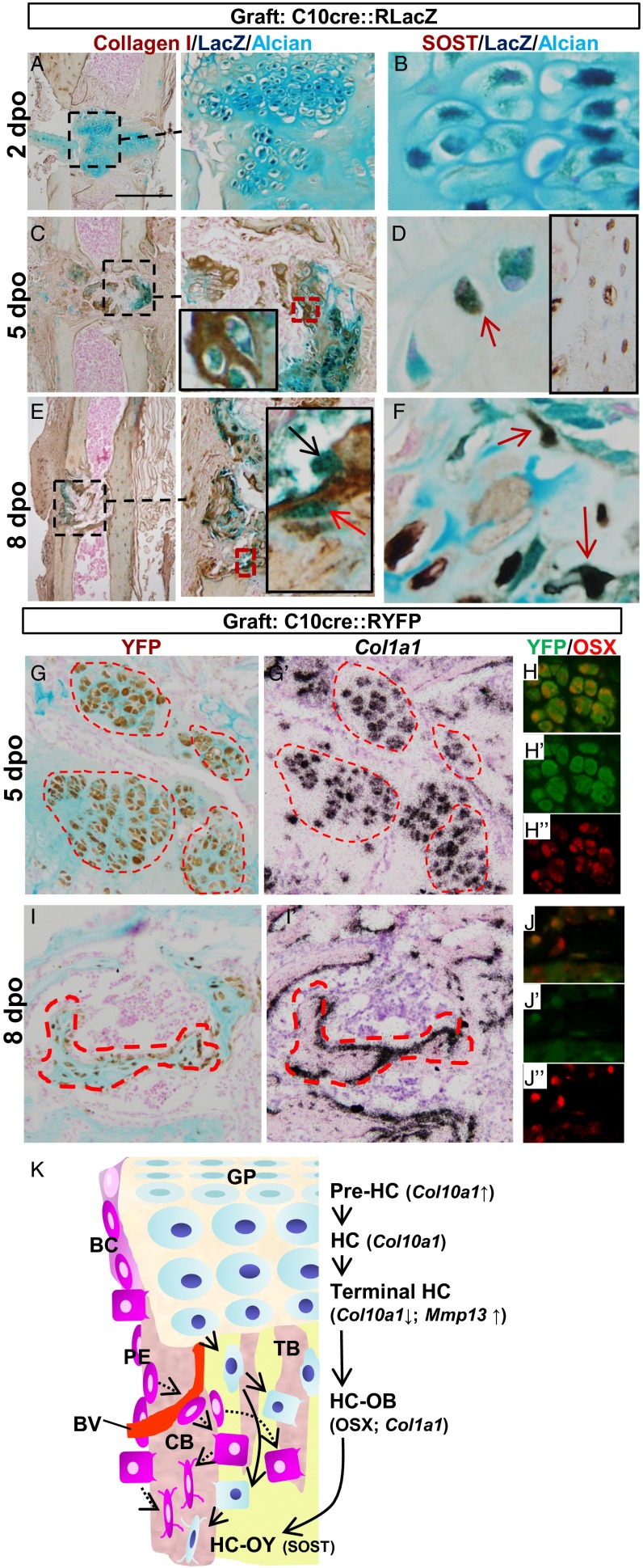
Fig. 5.
HC-derived osteoblasts and osteocytes contribute to bone repair and revised concept of osteoblast ontogeny in endochondral bone. (A–F) Fate of LacZ-tagged HCs in grafts of P10 C10cre::RLacZ hypertrophic cartilage inserted into the injury sites in tibia of P3m adult females. Tibiae were analyzed for indicated markers 2, 5, and 8 dpo. Alcian blue and collagen I immunostaining marks cartilage and bone matrix, respectively. LacZ+ (blue) osteoblasts (black arrow) and osteocytes (red arrows) of graft origin were identified in the bone repair site. (G–J) Similar to A–F, graft hypertrophic cartilages from C10cre::RYFP mice were inserted into the bone injury site. YFP+ cells expressing Col1a1 and OSX were detected at 5 and 8 dpo. (K) A model for the ontogeny of osteoblasts in endochondral bone. Sources of osteoblasts are direct differentiation from periosteal mesenchymal cells to form cortical bone (CB), perichondrium-derived osteoblast progenitors accompanying vascular invasion of the POC, and HC transition to osteoblast lineages. BC, bone collar; BV, blood vessel; GP, growth plate; OB, osteoblast; OY, osteocyte; PE, periosteum; TB, trabecular bone.

