
Fig. 5.
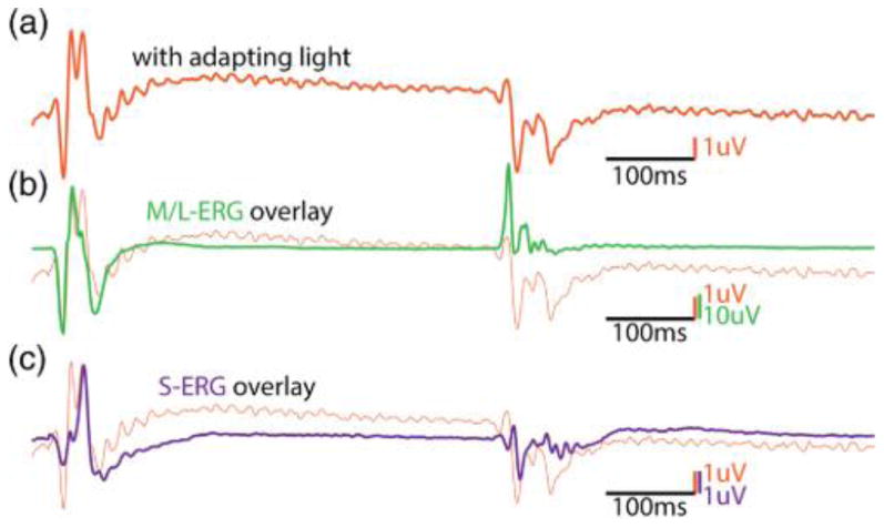
(a) Contains an ERG that was elicited in response to a 420 nm wavelength LED in the presence of a ∼30,000 td594 nm amber suppressing light. At light onset, two distinct peaks can be observed. (b) Overlays the L/M ERG arbitrarily scaled from the same animal. The timing of the peak lines up with the first peak, indicating this is +(L+M) contamination. (c) Overlays the pure S-cone ERG elicited from the same animal using the technique described in this paper. The S-cone peak is aligned with the second peak, indicating this is a pure S-cone mediated signal.