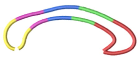
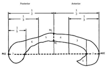
Table 2. Corpus callosum sections according to the topography of cerebral connected areas.
| Corpus callosum sections | Hemispheric connected areas |
|
|---|---|---|
Current Study
|
Witelson's subdivision
|
|
| 1-33 (red) Section A | Rostrum, Genu, Rostral body (regions 1, 2, 3) | Prefrontal, premotor, supplementary motor |
| 34-50 (green) Section B | Anterior mid-body (region 4) | Primary motor |
| 51-67 (blue) Section C | Posterior mid-body (region 5) | Somesthetic, posterior parietal |
| 68-81 (purple) Section D | Isthmus (region 6) | Superior temporal, posterior parietal |
| 82-100 (yellow) Section E | Splenium (region 7) | Inferior temporal and occipital |
Correspondence between the 100 callosal points clustered in five sections (from A to E), as were used in the present work, compared to the “classical” Witelson's subdivions, according to interhemispheric connected cortical areas.
