
FIGURE 4.
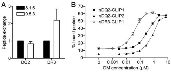
DQ2 displays reduced DM mediated peptide exchange compared to DR3 (A) Lysates of 8.1.6 and 9.5.3 B-LCLs were added to anti-DQ2 (2.12.E11) or anti-DR (L243) coated plates, followed by 48 h incubation with high affinity biotinylated indicator peptides (EPRAPWIEQEGPEYW for DQ2 and KTIAYDEEARR for DR3). The peptide exchange of DQ2 or DR3 from 8.1.6 (wt) is assigned a value of 1. Mean and SD from four independent experiments are shown. (B) Peptide exchange of cleaved sDQ2-CLIP1, sDQ2-CLIP2 and sDR3-CLIP1 (1.5 μM) with high affinity indicator peptides (1–5 nM of KPLLIIAEDVEGEY for DQ2 and KTIAYDEEARR for DR3) were measured in the absence (0 DM) or presence of an increasing amount of DM (0.001–7.2 μM) using the fluid-phase peptide binding assay. The complexes of DQ2 or DR3 with indicator peptides were used as a measure of peptide exchange. The negative control (without DQ2/DR3) did not give any signal (not shown). The mean and range of two independent experiments is shown.