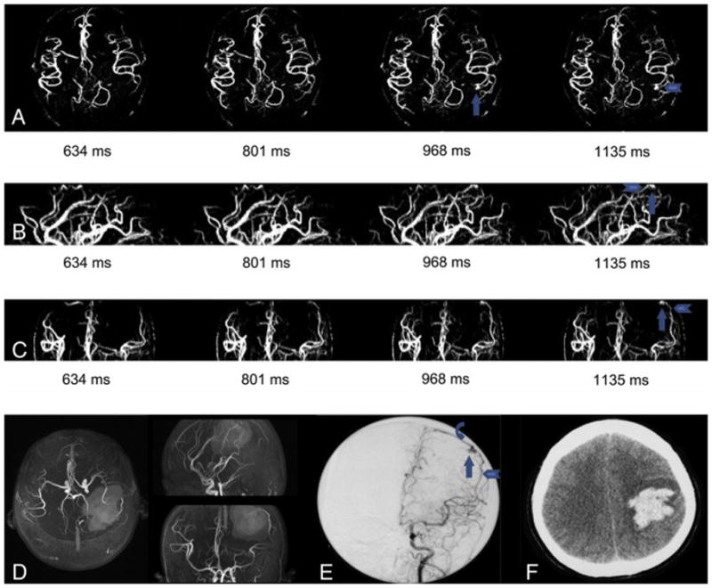
Figure 3.

Patient No. 7 male, 40 years old. He experienced spontaneous intracranial hemorrhage (d) and received conservative treatment in his regional hospital 3 month before he was admitted into Tiantan Hospital. dMRA didn’t detect any lesion (a) and TOF seemed to detect a frontal lesion (b, arrowhead). DSA demonstrated a frontal AVM with slow blood flow (c, arrow) and the lesion was lightly stained and should be identified with efforts. We compared DSA and TOF carefully and found that the lesion showed on TOF was actually caused by high signal intensity caused by methemoglobin and the real AVM entity located laterally to the hematoma. This was verified by operation.
