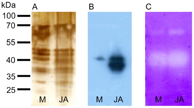
Figure 10. Protein pattern, Western blot and zymogram in response to external application of jasmonic acid.

Silver-stained SDS-polyacrylamide gel containing proteins in D. muscipula digestive fluid after mechanical stimulation (M) and external application of jasmonic acid (JA), (A). The same amount of proteins was electrophoresed in 12% (v/v) SDS-polyacrylamide gel and subjected to Western blot analysis using antibodies against dionain-1 (B). Detection of protease activity in 12% (v/v) SDS-polyacrylamide gel with casein as a substrate (C). The clear bands against background indicate protease activity. Representative gels at least from 4 repetitions are shown.
