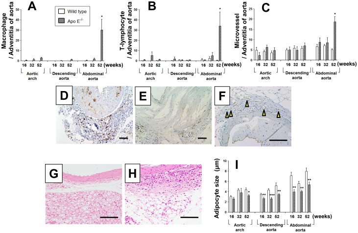
Figure 2. Impact of age on adventitia inflammatory cells, microvessels, and perivascular adipocytes.
(A–C) Numbers of F4/80-positive macrophages (A), CD3-positive T-lymphocytes (B), and CD31-positive microvessels (C) in the adventitia of the aorta at each age and region in apo E−/− and wild-type mice. (D–F) Representative images of F4/80 (D), CD3 (E), and CD31 (F) immunostaining in the abdominal aorta of apo E−/− mice at 52 weeks. Arrows indicate CD31-positive microvessels in the adventitia. (G, H) Representative hematoxylin–eosin staining of perivascular adipose tissue in the descending aorta (G) and abdominal aorta (H) of 52-week-old apo E−/− mice. (I) Diameter of adipocytes surrounding the aortic arch, descending aorta, and abdominal aorta. Data are expressed as mean ± SEM [16 weeks: wild-type (n = 9), apo E−/− mice (n = 5–6); 32 weeks: wild-type (n = 5–7), apo E−/− mice (n = 6–7); 52 weeks: wild-type (n = 10), apo E−/− mice (n = 9–11)]. *p<0.05, **p<0.01 vs. wild-type mice. Scale bar, 100 µm.

