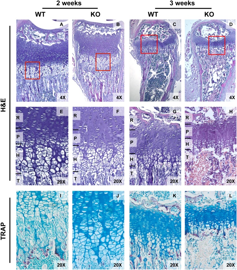
Figure 3. Progressive bone marrow hypoplasia with defective ossification, loss of trabecular bone and thinning of cortical bone is seen by 3 and 4 weeks in Tle4 null mice.
A–D. Hematoxylin and Eosin (H&E) staining of tibiae of 3 and 4 week old WT and Tle4 null (KO) mice demonstrate multiple abnormalities in Tle4 null mice including progressive pancytopenia of the bone marrow (BM), loss of trabecular bone (T), and thinning of the cortical bone layer (C). A higher power view shows a thinner proximal tibial growth plate in Tle4 null mice with a decrease in thickness of the resting (R), proliferative (P), and hypertrophic (H) zones and near complete loss of the trabeculae. E–H. Tartrate-resistant acid phosphatase (TRAP) staining (pink) demonstrates osteoclasts clustering under the hypertrophic zone at the boundary of the bone marrow cavity in Tle4 null mice at 3 and 4 weeks of age.

