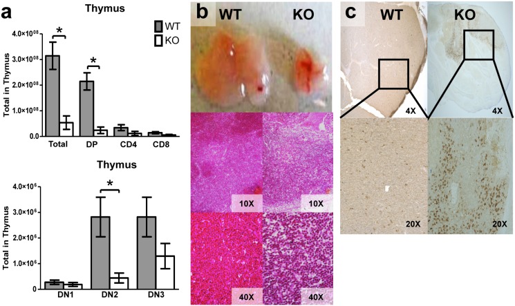
Figure 6. Tle4 null mice develop thymic atrophy with a block in T-cell differentiation.
(a) There is a dramatic decrease in total thymocytes in 3 week old Tle4 null mice as compared to wild-type littermates. The majority of this decrease is due to loss of double positive CD4+CD8+ cells. Within the double negative (DN) T progenitor populations there appears to be a block between DN1 (CD44+CD25−) and DN2 (CD44+CD25+) with a significant decrement in DN2 cells and an insignificant decrease in DN3 (CD44−, CD25+) cells. (n = 3–4 per genotype; mean +/−SEM; *: P<.05). (b) The thymus of 3 week old Tle4 null mice is atrophied with a loss in the demarcation between cortex and medulla as seen by H&E. (c) TUNEL staining demonstrates thymic apoptosis in Tle4 mice.

