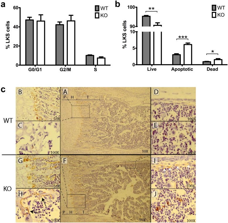
Figure 8. The decrease in LKS cells in Tle4 null mice is due to an increase in apoptosis and cell death rather than a decrease in proliferation and is accompanied by abnormalities of the bone marrow stroma.
(a) LKS cells isolated from the bone marrow of two week old mice show no difference in cell cycle distribution. (b) There is however an increase in apoptotic and dead LKS cells in the Tle4 null mice. (c) TUNEL staining of the growth plate of the femur in two week old mice marks the normal zone of cell death between the hypertrophic (H) layer and forming trabecular (T) bone (A, B) with an increase in staining in Tle4 null mice (F, G). Lacunae in the epiphysis are lined with periosteal cells undergoing apoptosis in Tle4 null mice (H), but was not seen in wild type (WT) littermates. Similar periosteal cells undergoing apoptosis and stained by TUNEL are seen under the cortex of diaphyseal bone in Tle4 null mice (I) but absent in wild-type mice (D). An increase in TUNEL staining is also observed in cells of the bone marrow in Tle4 null mice (J) as compared to wild-type bone marrow (E).

