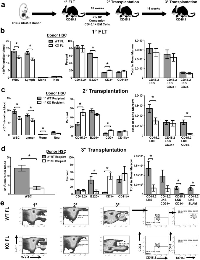
Figure 12. Tle4 null fetal liver HSCs have impaired B cell development and exhaust with serial transplantation.
(a) Schematic of serial FL transplantation experimental design. (b) Peripheral blood and LKS analysis of FL transplant recipients 16 weeks after transplant revealed peripheral leukopenia (WBC) and specifically B cell (B220+) lymphopenia in the absence of Tle4 with no difference in LKS, LKS, CD34+, LKS CD34− populations (n = 10 per genotype for blood analysis, n = 6–7 per genotype for LKS analysis, ***: P<.0001). (c) Peripheral blood and LKS analysis 16 weeks after secondary transplantation also indicates leukopenia and lymphopenia in Tle4 null cells, but at this time also a significant decrease in LKS and LKS, CD34− populations (n = 5 KO recipients, n = 10 WT recipients for blood analysis, n = 5 mice per group for LKS analysis, *: P<.05 **: P<.001). (d) Peripheral blood analysis and LKS analysis 16 weeks after tertiary transplantation again showed leukopenia, B-cell lymphopenia, and a profound decrease in all HSC containing populations (n = 4 per genotype for blood and LKS analysis, *: P<.05, **: P<.01, ***: P<.001). (e) Representative flow cytometry plots showing progressive loss of Tle4 null HSCs over successive transplantation.

