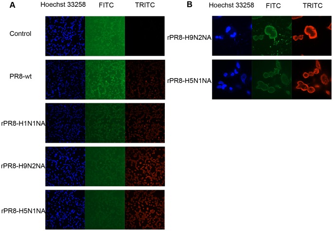
Figure 5. UBE2NL protein in virus-infected or mock-infected MDCK cells at 6 h p.i..
MDCK cells were infected with the viruses at MOI of 0.1 in the presence of 1 µg/ml TPCK-trypsin. After adsorption for 1 h at 37°C, the inocula were removed and the cultures were incubated for 6 h at 37°C in the maintenance media. Then, the cells were processed for indirect immunofluorescence assay, and the infected cells were detected with polyclonal antisera to UBE2NL protein and NP protein. (A) The fluorescence images (10×) of the infected and mock-infected cells at 6 h p.i. The FITC-fluorescence signal was expressed as UBE2NL protein and TRITC-fluorescence signal was expressed as the infected cells. (B) The fluorescence images (60×) of the cells infected by rPR8-H9N2NA or rPR8-H5N1NA viruses.

