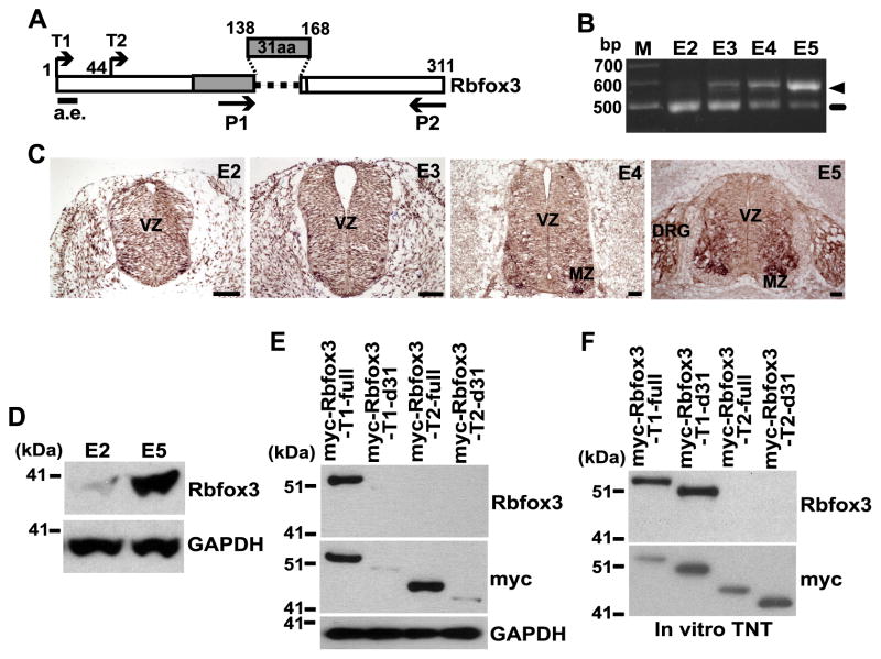
Fig. 1.
The Rbfox3-d31 protein is susceptible to degradation in cells. (A) Diagram of chicken Rbfox3 isoforms. The 31aa are encoded by the alternative exon. The N-terminal residue for the T1 and T2 constructs are shown. P1 and P2 indicate the primers for RT-PCR. a.e. indicates the epitope region for the anti-Rbfox3 antibody. Numbers represent amino acids. RNA-recognition motif (RRM) is indicated by gray bar. (B) Differential mRNA expression of Rbfox3 isoforms during chicken embryo development. RNA extracted from the embryo at the indicated embryonic day was analyzed by RT-PCR using the P1 and P2 primers indicated in panel A. Ethidium bromide stained agarose gel following electrophoresis of Rbfox3-PCR products is shown. The arrow head and line indicate the full-length Rbfox3 and Rbfox3-d31 mRNAs, respectively. M, size marker. (C) Rbfox3 mRNA expression in the developing chicken neural tube. In situ hybridization detecting Rbfox3 mRNA is shown. In situ-positive signals are shown as dark brown staining. Scale bars 50 μm. VZ, ventral zone; MZ, marginal zone; DRG, dorsal root ganglion. (D) Expression of endogenous Rbfox3 protein during development. Chicken embryo extracts harvested at the E2 and E5 were subjected to immunoblotting for the Rbfox3 and GAPDH proteins. (E) Differential protein stability of Rbfox3 isoforms in intact cells. Expression constructs indicated at the top of the blots were transfected into HEK-293 cells. Cell lysates were subjected to immunoblotting for the indicated proteins. (F) Similar protein stability of Rbfox3 isoforms in a cell-free system. Myc-Rbfox3 protein was synthesized in vitro from the indicated constructs at the top of the blots using the TNT Coupled Reticulocyte Lysate System followed by immunoblotting for the indicated proteins.

