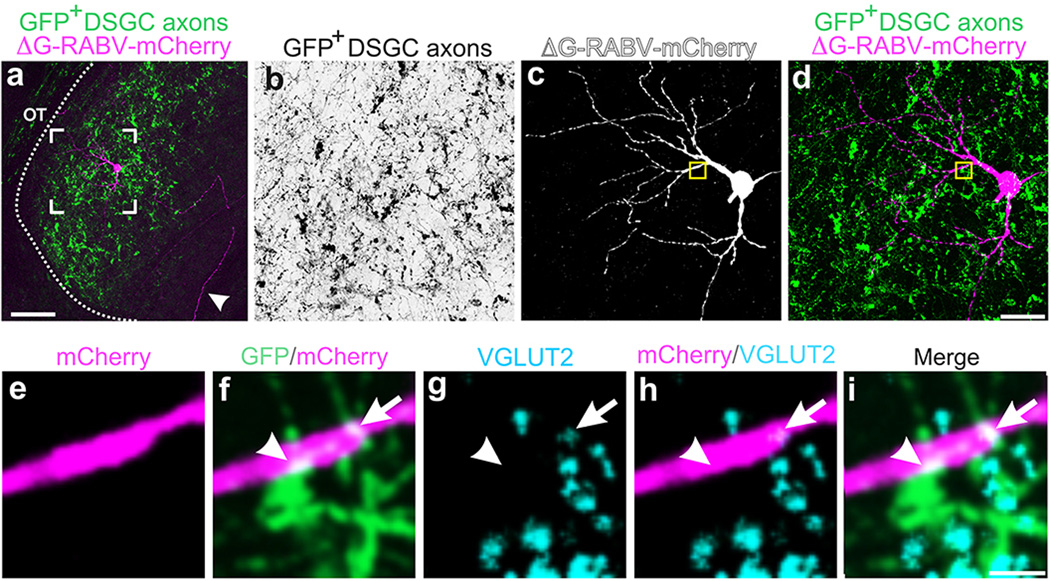
Extended Data Figure 5. Putative sites of contact between DSGC axons and a dLGN neuron retrogradely infected from superficial V1.
a–i, GFP+ On-Off DSGC axons (green in all panels except black in b) and mCherry+ dLGN relay neuron (magenta in all panels except white in c) infected by injection to superficial V1. Framed region in a is shown at higher magnification in b–d. Arrowhead (a): thalamocortical axon of mCherry+ dLGN cell. Scale in a, 50 µm. Yellow boxed region in c,d, is shown at higher magnification in e–i. Scale in d, 15 µm. e–i, Some DSGC axon-dendrite contacts contain VGLUT2 (blue). f–i, arrowhead: site of GFP/mCherry co-localization that does not contain VGLUT2. Arrow: GFP/mCherry/VGLUT2+ contact.

