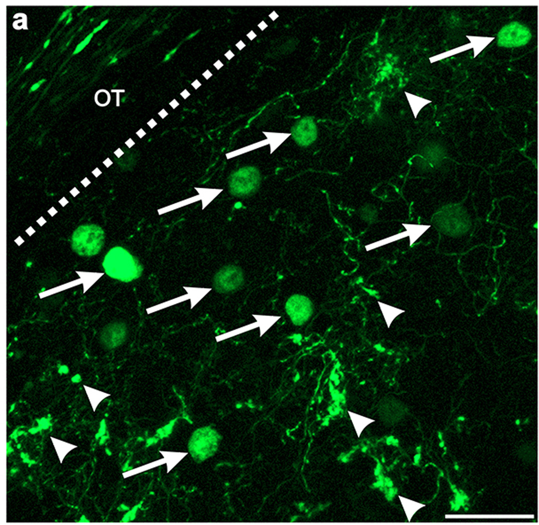
Extended Data Figure 6. The axons of GFP+ On-Off DSGCs and dLGN neurons infected with AAV2-Glyco-hGFP can be distinguished on the basis of their cellular localization.
High magnification view of DSGC-RZ in mouse with GFP + posterior-tuned On-Off DSGCs that was injected 14 days prior with AAV2-Glyco-hGFP. Glyco-hGFP+ neurons have nuclear GFP labeling (arrows) whereas DSGCs have GFP in axon terminals (arrowheads). Scale, 50 μm.

