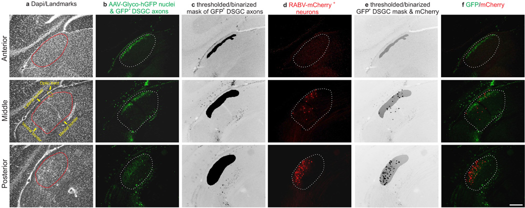
Extended Data Figure 4. Analysis of dLGN neurons retrogradely infected from superficial V1.
a–f, Example serial sections of anterior, middle and posterior portions of dLGN in a mouse with GFP expressing On-Off DSGC axons that was injected with ΔG-RABV-mCherry in superficial layers of V1. a, dapi to show cytoarchitectural landmarks and dLGN borders. b, GFP+ DSGC axons and AAV-Glyco-hGFP infected cell bodies (see main Fig. 4 and text). c, Mask of GFP+ DSGC axons (Methods). d, ΔG-RABV-mCherry+ dLGN relay neurons. e, GFP+ DSGC axon mask superimposed with mCherry signal; this was used to determine colocalization. f, mCherry and hGFP signals merged. Scale, 200 μm.

