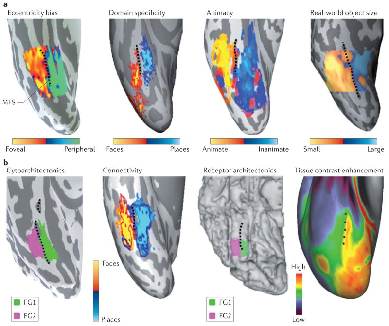
Figure 4. Linking anatomical features to large-scale functional maps in the ventral temporal cortex.
a | The mid-fusiform sulcus (MFS) predicts transitions in many large-scale functional maps in the ventral temporal cortex (VTC). Lateral–medial functional transitions in the eccentricity bias map15 (based on data from REF. 17), the domain-specificity map (based on data from REF. 33), the animacy map (based on data from REF. 116) and the real-world object-size map (based on data from REF. 27 and T. Konkle, personal communication) are all aligned to the MFS (shown by the dashed black line). Each panel shows a representative inflated right hemisphere from an individual subject, with the exception of the domain-specificity map, which was generated from ten subjects. b | The MFS predicts transitions of anatomical features of the VTC. Lateral–medial anatomical transitions in cytoarchitecture, in white-matter connectivity, in the density of muscarinic acetylcholine receptor type 3 and in tissue contrast enhancement (which is thought to be related to myelin content) are each aligned to the MFS. Each panel shows a representative right hemisphere, with the exception of the tissue contrast enhancement map, which is generated from 196 subjects from the Human Connectome Project (based on data from REFS 122,123). FG, fusiform gyrus. The cytoarchitecture panel is based on data from REF. 17. The connectivity panel is based on data from REF. 30 and Z. M. Saygin, personal communication. The receptor architectonics panel is based on data from REF. 29. Receptor architectonics panel courtesy of J. Caspers, Institute of Neuroscience and Medicine (INM-1), Research Centre Jülich, Germany. Tissue contrast enhancement panel courtesy of M. F. Glasser and D. C. Van Essen, Washington University, St Louis, Missouri, USA.

