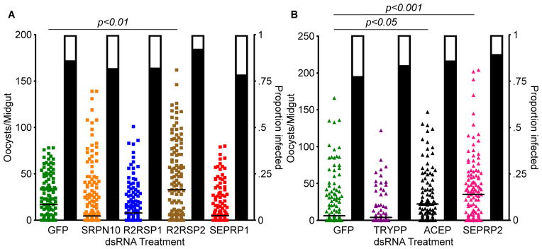
Figure 4. P. falciparum infection intensity following RNAi knockdown of protease genes.

The number of oocysts per midgut of wild-type A. stephensi following RNAi-mediated depletion of: A) green fluorescent protein (GFP), serpin 10 (SRPN10), Rel2-responsive serine protease 1 (R2RSP1), Rel2-responsive serine protease 2 (R2RSP2), or serine protease precursor 1 (SEPRP1); and B) trypsin precursor (TRYPP), anigiotensin-converting enzyme precurser (ACEP), or serine protease precursor 2 (SEPRP2). Silencing of R2RSP2, ACEP, and SEPRP2 all led to significant increases in the number of oocysts per midgut. Each circle represents a single midgut, and horizontal black bars represent the median of the sample. Significance was determined by a Kruskal-Wallis test followed by Dunn’s post-hoc test to compare immune-depleted mosquitoes to GFP controls. Significance was assessed at α=0.05. Supplementary data for this figure is given in table S2.
