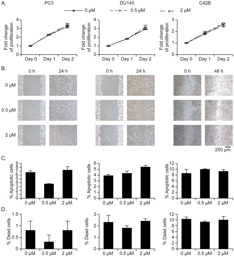
Figure 5. Effects of DiD on cell proliferation, migration, and apoptosis.
To determine possible adverse effects of DiD on cell function, PCa cells (PC3, DU145, C42B) were stained with DiD (0.5 μM and 2 μM), and a selection of functional assays were performed. (A) Cell proliferation was measured by MTS assay. (B) Cell migration was assessed using a wound-healing assay. Original magnification, 10x / 0.25 Ph1 ADL. Scale bars: 250 μm. After 2 days of DiD staining, (C) apoptosis was analyzed by flow cytometry with Annexin V staining and (D) the number of dead cells was counted by trypan blue staining. Data are presented as mean ± standard deviation from triplicate determinations.

