
Figure 3. MMP-9 inhibition abrogates endothelial dysfunction induced by NETs.
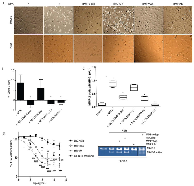
A. HUVECs and HAECs were incubated for 3 h in the absence or presence of NETs (+), MMP-9-depleted (MMP-9 dep)-NETs, histone H2A-depleted (H2A dep)-NETs, NETs plus neutralizing anti-MMP-9 (MMP-9 Ab) or NETs plus MMP inhibitor (MMP inh). Images display HUVEC morphological changes induced by NETs. B. Apoptotic ECs were quantified by FACS. Results are adjusted to background levels (no NETs) and represent mean ± SEM of six independent experiments. C. Zymography analysis performed on supernatants after 3 h incubation. MMP-2 (representative image, bottom) was quantified by densitometry. Results are presented active MMP-2/pro-MMP-2 ratio and expressed as median AUs ± SEM. D Improvement in aortic endothelium-dependent vasorelaxation after incubation with lupus LDG NETs with and without neutralizing anti-MMP-9 (MMP-9 Ab) or MMP inhibitor (MMP inh). Results represent mean ± SEM of two experiments. *p< 0.05, **p< 0.005, ***p<0.001, #### p< 0.0001 for Ctrl vs lupus LDG comparison; acetylcholine (Ach).