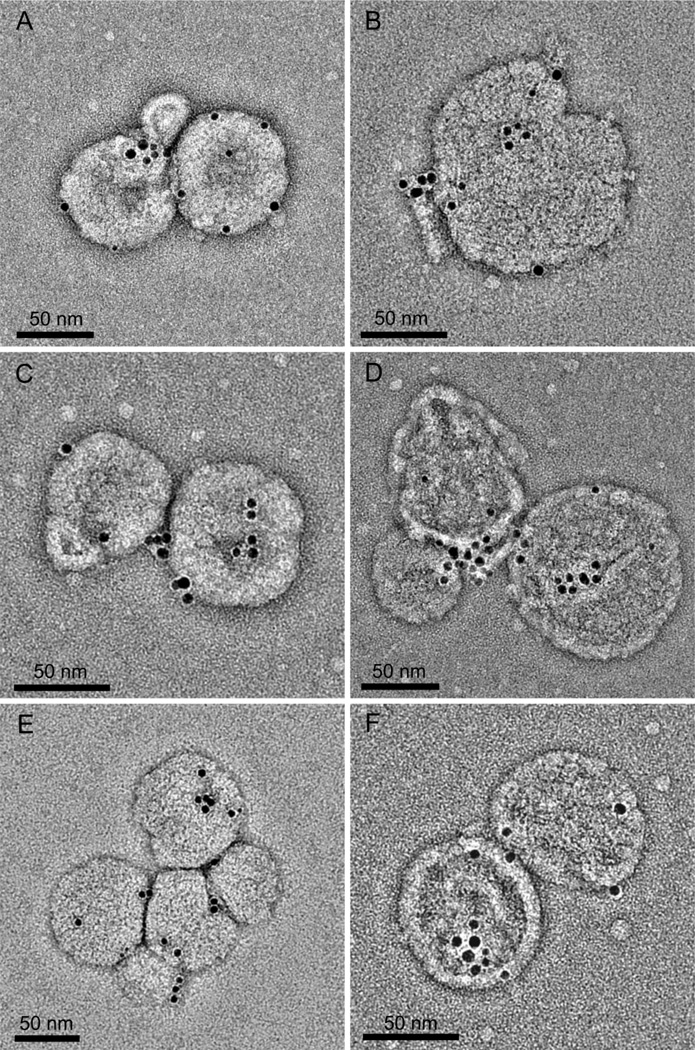
Figure 2. Identification and membrane localisation of cytbc1 complexes.
A–F. Negatively stained whole chromatophores with cytbc1 complexes labelled with gold NTA-Nanogold®. The edge-to-edge separation of pairs of gold beads is 2.4 ± 0.5 (S.D.) nm n=118, compatible with the structure of the Rba. sphaeroides cytbc1 dimer complex12,13 and also the surface shell of the Ni-NTA nanoparticle.

