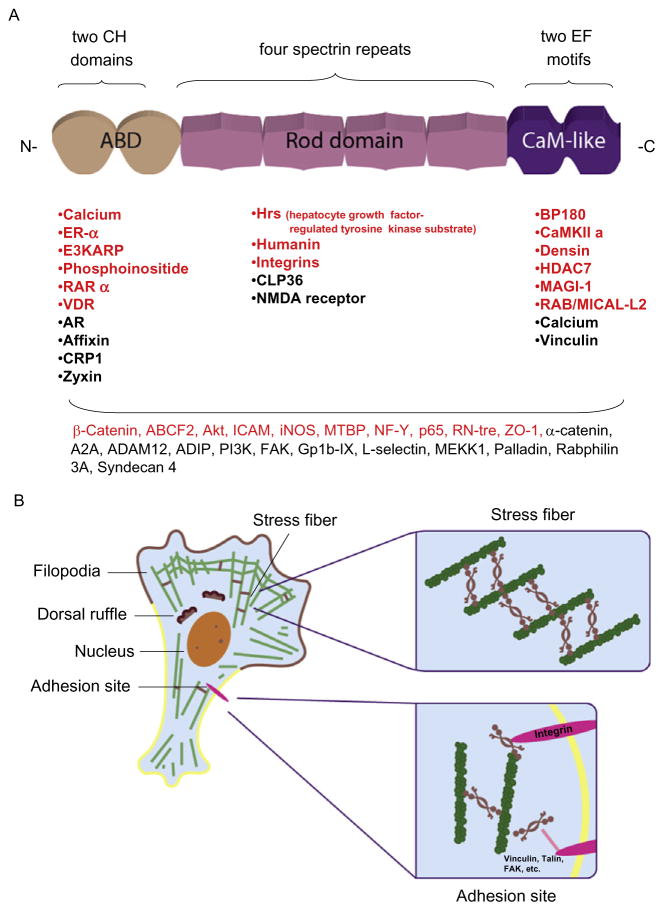
Figure 13.1.
(A) A schematic representation of the structural domains of ACTN. The N-terminal CH repeat constitutes a functional actin-binding domain (ABD), which can also bind to phosphoinositide and calcium. Following the ABD, four SRs form a rod-shaped structure that connects to the EF motif-integrated CaM-like domain. A list of ACTN binding partners is also shown under the corresponding domains. Among these binding partners, the proteins annotated in red are validated ACTN4 binding partners. Shown at the bottom are ACTN binding partners of which the interaction domains were not mapped. (B) A cartoon depicting a moving cell demonstrating the detailed localization of ACTN4 in stress fiber and adhesion sites. F-actin is shown in green and ACTN4 is shown as a brown line or a twisted cherry-like shape. Inside the cells, ACTN4 cross-links with F-actin to form stress fibers or locates in other motile apparatus components or in the nucleus. Close to the focal adhesion sites, ACTN4 directly links integrins with F-actin or other adhesion molecules, such as vinculin or talin.

