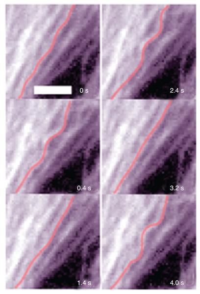
Figure 5.
Microtubules visualized over time in a beating cardiac muscle cell expressing green fluorescent protein-tubulin. Whenever the cell contracts, the microtubule (highlighted in red) buckles indicating that it resists contraction of the cytoskeletal actin network. Scale bar = 3 μm. Reproduced from reference 12 with permission form Brang-wynne et al.

