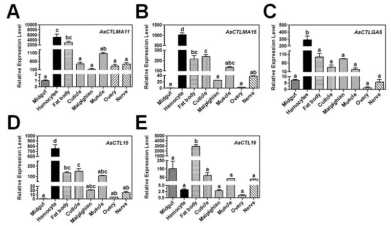
Fig. 2. Tissue distribution of AsCTLs in Ar. subalbatus females.

Midgut, hemocytes, fat body, cuticle, Malpighian tubule, muscle, ovary and nerve were dissected from female mosquitoes (4–5 days old) and used for preparation of total RNAs. Expressions of AsCTLs transcripts were determined by real-time PCR. AsRPL9 gene was used as an internal control gene. Each bar represents the mean of three individual measurements ± SEM. Identical letters are not significant difference (p>0.05), while different letters indicate significant difference (p<0.05) determined by one way ANOVA followed by a Tukey’s multiple comparison test among different tissues.
