
Figure 1.
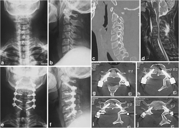
Radiographic studies of the cervical defects and performed procedures. (a–c) Preoperative anteroposterior images of vertebral fracture in cervical vertebrae C2 and C3. (d) Spinal stenosis between C3 and C4, and between C4 and C5 are shown with preoperative magnetic resonance imaging; spinal cord deformation, posterior ligament complex injury, and hematoma anterior to vertebrae are revealed between C2 and C5. (e–f) Open-door posterior laminoplasty and transpedicle internal fixation at C2–C5. (g–j) Transpedicle screws were embedded into the pedicle.