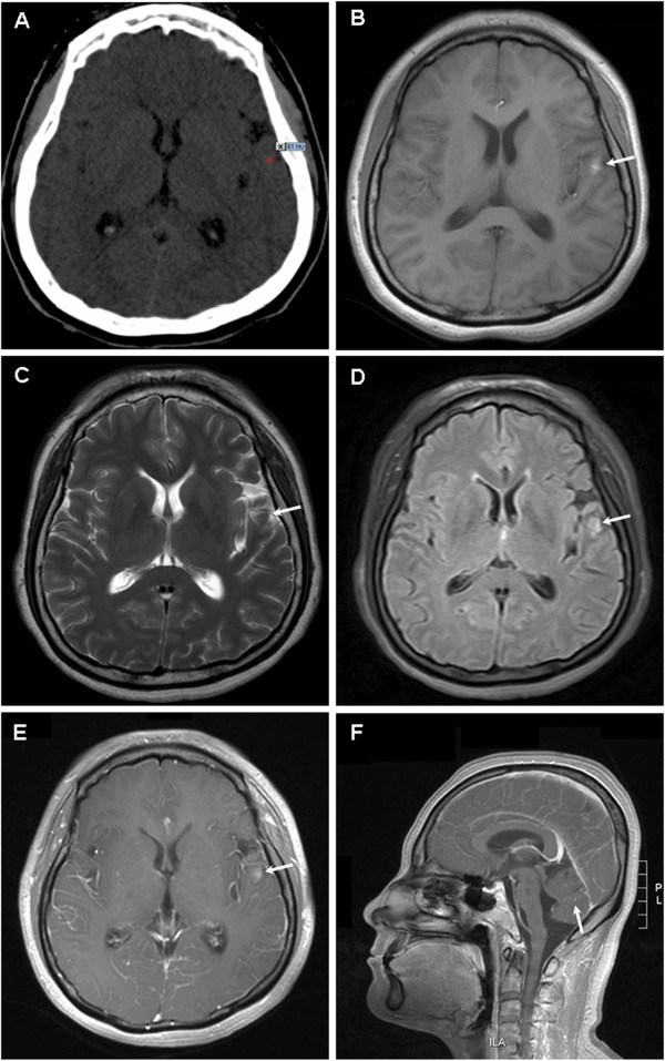
Figure 1.

Imaging findings for brain. (A) Computed tomography image showing an isodense or slightly hyperdense lesion in the left temporal lobe (CT value: 45 to 61). Magnetic resonance image showing a 6-mm abnormal signal in the left temporal lobe, which was hyperintense on T1-weighted images (B), hypointense on T2-weighted images (C), slightly hyperintense on fluid-attenuated inversion recovery images (D), and mild enhancement with gadolinium contrast (E). Punctate and linear enhancement in the sulci and gyri of the cerebellum can be observed (F).
