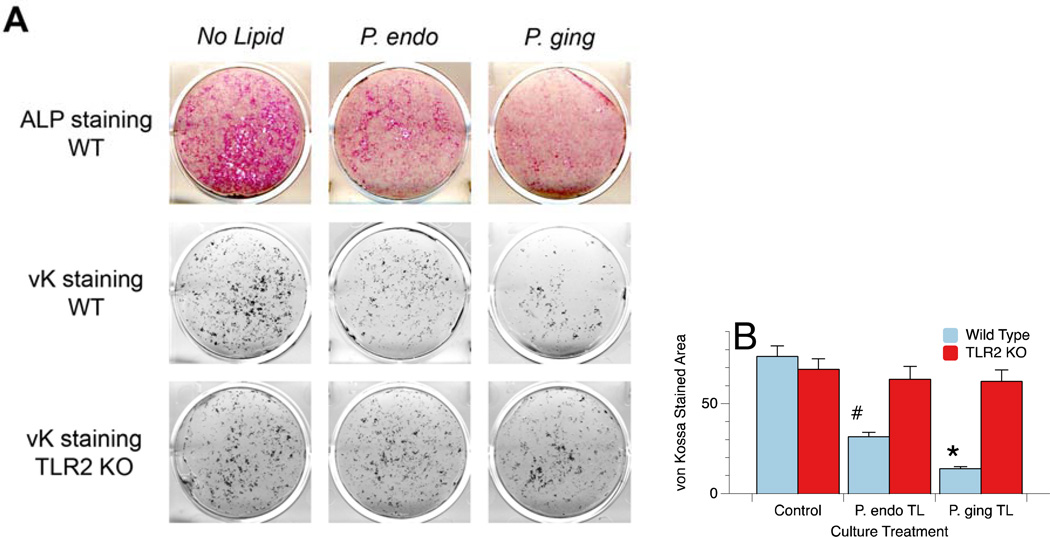
Figure 2.
Wild type (WT) and TLR2−/− (KO) osteoblasts treated with total lipid extracts from P. endodontalis or P. gingivalis and stained for alkaline phosphatase (ALP) or mineralization nodule formation by von Kossa (vK) staining (Figure 2A). Osteoblasts were treated with 1.2µg/ml of P. endodontalis or P. gingivalis total lipid extracts for days 7 through 21. Note that the total lipid extract of both P. endodontalis (P. endo) and P. gingivalis (P. ging) inhibit alkaline phosphatase expression and von Kossa stained mineral deposition in vitro, and the mineral deposition may be TLR2 dependent as has been previously reported for P. gingivalis total lipids (16). Quantitation of the von Kossa (vK) stained mineral deposits was accomplished using ImageJ processing and revealed that in WT cultures, the area of vK staining was significantly less in both P. endo total lipid (TL, #p<0.002) and P. ging TL (*p<0.001) treated osteoblasts cultures (Figure 2B). In contrast, mineral deposition was not significantly affected in osteoblasts derived from TLR2−/− animals.

