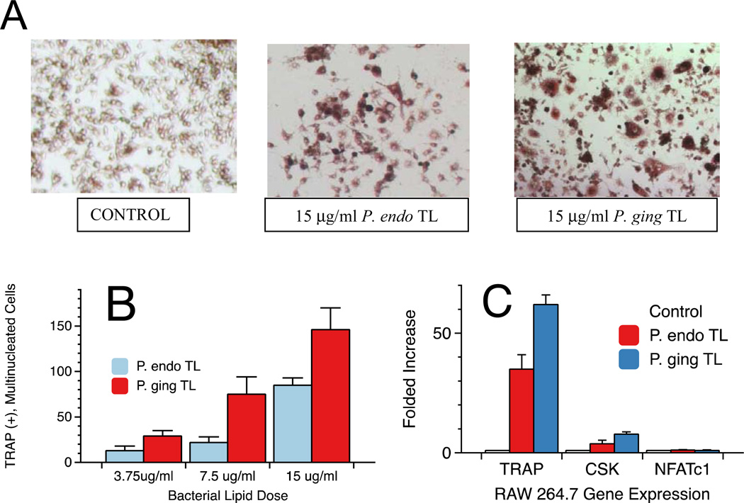
Figure 3.
Osteoclast formation from RAW 264.7 cells following exposure to total lipids of either P. endodontalis or P. gingivalis. Bacterial lipid effects on the formation of TRAP positive (+), multinucleated cells in cultures were evaluated following treatment with total lipid extracts from either P. endodontalis or P. gingivalis. Bacterial lipids at the indicated concentrations were sonicated in medium before addition to cell cultures. Cells were exposed to lipids for 7 days before staining and microscopic visualization of osteoclasts. Osteoclast formation varied depending on lipid treatment as shown in Figure 3A. TRAP(+), multinucleated giant cells in culture wells were counted and averaged for three wells (Figure 3B). The values were expressed as the mean ± standard error (SE) of triplicate cultures. Control cultures showed no TRAP(+), multinucleated cells. By one factor ANOVA, the number of TRAP(+), multinucleated cells was significantly increased with higher doses of each total lipid preparation when compared with the respective lower doses (p<0.05). Osteoclast-specific gene expression was evaluated in RAW 264.7 cells exposed to 15 µg/ml of P. endodontalis or P. gingivalis total lipids cultured for seven days (Figure 3C). RNA was extracted as described in the Materials and Methods and gene expression quantified by RT-PCR. Each sample was assayed in triplicate.

