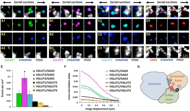
Figure 3.
Glutamate and GABA axon boutons in the DRN. (A–D) Array tomographic visualization across four ultrathin (70 nm) serial-sections immunolabeled against VGLUT1-3 (green, magenta, and light blue, respectively) or GAD2 (red) to identify glutamate and GABAergic boutons, respectively. These immunodetected axon boutons are often co-labeled for synapsin (blue), and in some cases associated with TPOH-labeled dendrites (white). Adapted from Soiza-Reilly et al. (2013) reprinted with permission. (E) Total density of axo-axonic-like arrangements of four different types of axon boutons labeled either for VGLUT1-3 or GAD2 together with synapsin. The most abundant pair of axon boutons contained VGLUT2 and GAD2 ones (*p < 0.001 vs. either VGLUT1 or VGLUT3). Only a small proportion of axoaxonic-like arrangements were found between different glutamatergic boutons (e.g., VGLUT1/VGLUT2, VGLUT1/VGLUT3, and VGLUT2/VGLUT3). Bars represent SEM. Adapted from Soiza-Reilly et al. (2013) reprinted with permission. (F) Cross-correlation analysis of pixels between four different types of axon boutons using the ImageJ plugin JACoP (Bolte and Cordelières, 2006). This analysis indicated a higher degree of colocalization of VGLUT2 axon terminals with GAD2 boutons in comparison to other glutamate axons. Specifically, when imaging channels were aligned (x = at 0 μm of displacement), the pixels co-labeled for VGLUT1-3 and synapsin showed the highest value of correlation with pixels co-labeled for GAD2 and synapsin. When imaging channels were displaced with respect to each other all of the correlation values decreased indicating the spatial proximity of all three glutamate axon types with GABA boutons (Soiza-Reilly et al., 2013). Cross-correlations between different glutamate axon types were very low. (G) Schematic illustration of the GABAergic presynaptic modulation of glutamate release in the synaptic triads of the DRN. Adjacent glutamate and GABA axon terminals are closely associated with a postsynaptic dendrite that receives a typical excitatory asymmetric synapse from the glutamatergic bouton. GABA boutons would have the capacity of presynaptically influencing glutamate release from terminals through a dual mechanism involving a rapid/transient GABA-A receptor-mediated facilitation, and a more delayed and sustained GABA-B receptor inhibition.

