
Fig. 3.
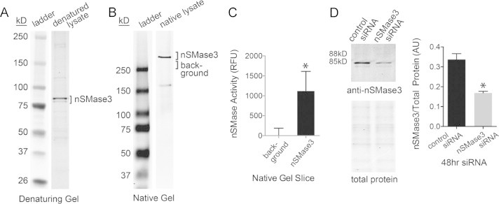
nSMase3 protein expression. (A) Western blot showing detection of a doublet using custom polyclonal nSMase3 antibodies. (B) Western blot under non-denaturing PAGE conditions shows migration of native nSMase3. (C) nSMase activity in non-denaturing gel slices of anti-nSMase3 reactive band compared to activity of non-specific gel slice (choline release, n=6), ⁎P<0.02. (D) siRNA specific for nSMase3 depletes the anti-nSMase3 reactive doublet. Sample volumes for western blot were adjusted for equal total protein, total protein was assessed by SDS-PAGE and simply blue staining (n=6, ⁎P<0.001).