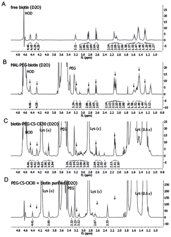
Figure 2.
1H-NMR spectrum of free and conjugated biotin. Free biotin (A), MAL-PEG-biotin (B), purified biotin-PEG-CK30 (C), and PEG-CK30 mixed with free biotin and then purified with ion exchange column (D) were dissolved in deuterium oxide and the 600 MHz NMR spectrum for each sample was obtained. Signals from PEG and polylysine were labeled in the figure. Signals from biotin were indicated by arrows.

