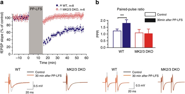
Figure 8. PP-LFS failed to induce mGluR-LTD in MK2/3 DKO mice.
(a) Application of PP-LFS (900 paired-pulses delivered at 1 Hz, with an inter-pulse interval of 50 ms) induced LTD of the field excitatory postsynaptic potential (fEPSP) in the CA1 region of WT hippocampus (blue square) but failed to induce LTD in DKO (red circle). Dots in the graphs represent the average slope for six consecutive fEPSPs; data are shown as mean±s.d. for number of experiments indicated. Averaged fEPSP (20 consecutive responses) obtained from one individual WT and DKO slices at baseline (black line) and 30 min post PP-LFS application (red line). (b) Pooled data showing the significant increase in paired-pulse ratio following PP-LFS in WT mice (control: 1.21±0.14; after LTD: 1.78±0.25), an effect that is absent in DKO mice (control: 1.07±0.24; after LTD: 1.01±0.33). Averaged fEPSP (20 consecutive events) obtained from one individual WT and DKO slices at baseline (black line) and 30 min post PP-LFS application (red line). Error bars indicate±s.d. and **P<0.01.

