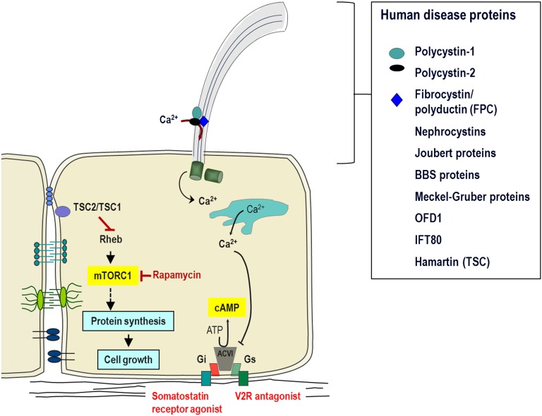
FIGURE 1.
The primary cilium and cystoproteins. The cilium concentrates and organizes a number of channels, receptors, and effectors, such as transcription factors and proteolytic fragments of cystoproteins. It therefore plays a critical role in transmitting information regarding the external milieu back into the cell and ultimately in regulating cellular and tubular differentiation and homeostasis. Cyst formation is characterized by deregulation of the balance between cell proliferation and differentiation. Cilia appear to play a role in maintaining this balance through sensing the extracellular milieu, responding to mechanical cues, and modulating different signaling cascades. Ciliary dysfunction contributes to increased intracellular accumulation of cAMP and activation of mammalian target of rapamycin (mTOR), features common to cystic epithelia in human and rodent models of renal cystic disease. Numerous groups have demonstrated that almost all cystoproteins, including polycystins; FPC; the nephrocystins; the Bardet Biedl syndrome (BBS) proteins; oral-facial-digital syndrome, type I (OFD1) protein; and the tuberous sclerosis type I (TSC1) protein Hamartin; all of these localize to the cilia/centrosome complex, providing compelling evidence that this complex is critical in the pathogenesis of renal cystic disease. mTORC1, mammalian target of rapamycin complex 1; V2R, V2 receptor.

